A cough lasting more than eight weeks does not automatically mean you need an invasive test. By the end, you will know when EBUS can investigate an abnormality beside the airways, what samples it can provide, which cough causes require different tests, and what to clarify before scheduling the procedure.
Key takeaways
- EBUS is considered when imaging shows lymph nodes or a tissue target.
- EBUS samples can help assess cancer, infection, and inflammatory disease.
- A normal EBUS does not rule out asthma, reflux, or airway sensitivity.
- Confirm the target, sedation plan, biopsy risks, and pathology process beforehand.
When does chronic cough evaluation reach the point of EBUS?
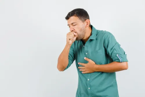
In adults, chronic cough means a cough lasting more than eight weeks. EBUS is not a routine first-line test; it becomes relevant when imaging identifies a target that may need tissue.
The initial work-up usually includes:
- A detailed cough history and chest examination
- Medication review, especially for ACE inhibitors
- Review of smoking, workplace, household, and other irritant exposure
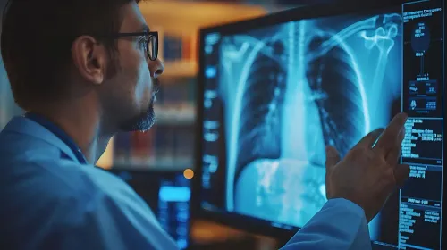
- Chest X-ray, followed by CT when indicated
- Spirometry and assessment for asthma or eosinophilic airway disease
- Evaluation for upper-airway cough syndrome and reflux
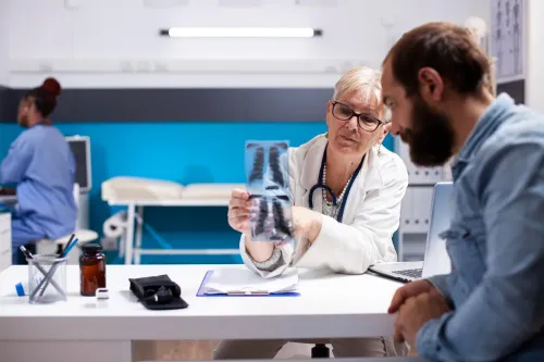
EBUS enters the discussion when CT shows an enlarged mediastinal or hilar lymph node, a mass beside the trachea or major bronchi, a peribronchial lesion, or another unexplained abnormality that could require sampling. The suspected diagnosis might include lung cancer spread, tuberculosis-related lymph-node disease, sarcoidosis, or another inflammatory condition.
A search for chronic cough in Mumbai or cough grant road does not itself indicate a need for EBUS. The imaging finding and suspected diagnosis determine whether the test can answer a useful question.
If the chest imaging is normal and the likely cause is asthma, nasal drainage, reflux, medication effect, or cough hypersensitivity, EBUS will not address the main problem.
What can EBUS check and what can the samples show?
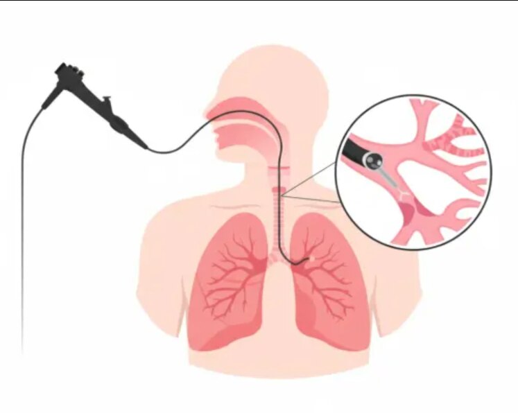
EBUS means endobronchial ultrasound: an ultrasound probe mounted on a bronchoscope images structures beside the airways. During chronic cough evaluation, convex-probe EBUS can locate and guide transbronchial needle aspiration of accessible mediastinal and hilar lymph nodes, selected central masses, and lesions next to the trachea or bronchi.
1. It can investigate suspected lung-cancer spread, sarcoidosis, tuberculosis-related lymph-node disease, and selected infections or inflammatory conditions when CT shows an abnormal target. EBUS is not simply observing the cough; it obtains material for diagnosis and, in cancer, sometimes staging.
2. The sample can undergo cytology, which examines individual cells, or histology, which examines tissue fragments. If infection is suspected, tell the bronchoscopy team before sampling so material is allocated for stains, bacterial or mycobacterial culture, and molecular tests. A specimen sent only for cytology may not provide enough material for microbiology or special testing.
3. The bronchoscope also inspects the airway lining. It can reveal narrowing, a visible tumour, mucus plugging, bleeding, foreign material, or an anatomical abnormality that ultrasound alone cannot show.
A search for “ebus bronchoscopy chronic cough in mumbai” or “ebus bronchoscopy in mumbai” does not determine suitability. The CT finding and suspected diagnosis do. A negative result also does not exclude every cancer, lymphoma, peripheral lesion, asthma, reflux-related cough, or cough hypersensitivity.
Which test answers the question EBUS cannot answer?
EBUS answers a tissue question about a reachable lymph node or central chest lesion; it does not explain every cause of chronic cough. A search for ebus bronchoscopy churchgate identifies a location, not the test that matches your imaging and suspected diagnosis.
| Test | Question it answers | Main limitation |
|---|---|---|
| Chest CT | What do the lung tissue, lymph nodes, and mediastinal structures look like? | It usually shows anatomy but does not provide tissue. |
| PET-CT | Which areas show increased metabolic activity, and where might disease have spread? | Inflammation and infection can also appear active, so tissue may still be needed. |
| Spirometry | Is airflow reduced, supporting asthma or obstructive lung disease? | It does not inspect lymph nodes or mediastinal masses. |
| Conventional bronchoscopy | Does the airway lining show narrowing, a visible tumour, bleeding, or mucus, and can washings or visible-airway biopsies be collected? | It does not provide EBUS ultrasound-guided sampling outside the airway wall. |
| EBUS bronchoscopy | Can a needle sample an accessible node or central lesion beside the trachea or major bronchi? | A negative result does not exclude every cancer, lymphoma, or peripheral lesion. |
| CT-guided needle biopsy | Can a selected lung or chest target reachable through the chest be sampled? | The route may not suit a central lesion or unsafe location. |
| Surgical mediastinoscopy | Is more tissue needed from the mediastinum through an operation? | It is more invasive than needle sampling. |
EBUS is not a good test for nasal disease, sinus drainage, reflux, vocal-cord dysfunction, most asthma, uncomplicated acute bronchitis, or many coughs with normal chest imaging. A negative result also does not exclude asthma, reflux-related cough, cough hypersensitivity, or all airway inflammation.
What happens before and during EBUS bronchoscopy?
1. Before EBUS bronchoscopy, the team reviews your CT images and suspected diagnosis, then checks allergies, sleep apnoea, heart or lung disease, and every medicine you take.
2. You receive fasting instructions and an individual plan for anticoagulants, antiplatelet drugs, and diabetes medicines. Do not stop any of these medicines without clinical instructions. Arrange an escort home because sedation or anaesthesia makes driving unsafe.
3. Consent should cover topical airway anaesthesia, moderate sedation or general anaesthesia, depending on the centre and procedure. It should also explain planned sampling, possible additional tests, alternatives, and what happens if the target cannot be safely reached. A search for “ebus bronchoscopy churchgate” does not replace this discussion.
4. During the procedure, the bronchoscope enters through your mouth or nose and passes into the airways. Ultrasound identifies a nearby lymph node or lesion, and the operator takes needle samples through the airway wall when the imaging and procedure plan show that tissue is needed.
5. Not every EBUS includes the same number or type of samples. Afterward, expect a sore throat, cough, small-volume bleeding, or fever. Uncommon risks include significant bleeding, infection, low oxygen, sedation effects, pneumothorax, and injury to nearby structures.
What should you confirm before choosing EBUS for chronic cough?
Choose EBUS only when CT shows a reachable abnormality and the tissue result will change treatment.
- Identify the exact target: an enlarged mediastinal or hilar lymph node, a mass beside the trachea or main bronchi, or a peribronchial lesion.
- Name the leading diagnosis: cancer, tuberculosis, sarcoidosis, lymphoma, or another inflammatory or granulomatous condition.
- Ask the team to divide the specimen appropriately for cytology, histology, bacterial or mycobacterial culture, stains, and molecular testing. A sample sent only for cytology may not support culture or special tests.
- Confirm that the target is accessible and that the planned number of needle passes can provide enough material, particularly if lymphoma is possible.
| Option | Best question it answers | Limitation |
|---|---|---|
| EBUS | Can a reachable central node or lesion provide diagnosis and cancer staging? | May not supply enough tissue for lymphoma |
| CT-guided biopsy | Can a peripheral lung or chest target be sampled through the chest? | Less suitable for central nodes or lesions beside airways |
| Conventional bronchoscopy | Is there an airway-surface lesion, narrowing, or visible tumour to sample? | Does not usually reach tissue outside the airway wall |
| Surgical mediastinoscopy | Is a larger node sample needed? | Requires an operation |
EBUS can diagnose and stage cancer in one procedure and avoid surgery, but surgery remains appropriate when the node is inaccessible or more tissue is needed. Interpret a result with imaging and the clinical picture; yield depends on lesion size, location, needle passes, technique, specimen handling, and disease type.
In a Mumbai consultation with Dr. Pujan Parikh, ask whether the scan shows a reachable target and whether the result will alter treatment—not whether a neighbourhood search term proves suitability.
Frequently asked questions
When does chronic cough evaluation reach the point of EBUS?
EBUS becomes relevant after chronic cough assessment and chest imaging identify enlarged lymph nodes, a central lung lesion, or another target near the airways that requires tissue. It is not a routine first-line test for every cough lasting more than eight weeks.
What can EBUS check and what can the samples show?
EBUS can examine structures beside the airways, including mediastinal and hilar lymph nodes, and collect needle samples. Pathology and microbiology can help assess cancer, tuberculosis and other infections, sarcoidosis, and selected inflammatory conditions.
Which test answers the question EBUS cannot answer?
The right alternative depends on the suspected cause. Spirometry with bronchodilator testing helps assess asthma or airflow obstruction; reflux evaluation, allergy assessment, sputum tests, or standard bronchoscopy may answer other questions that lymph-node sampling cannot.
What should you confirm before choosing EBUS for chronic cough?
Confirm the exact imaging target, whether needle sampling is likely to change treatment, sedation or anaesthesia arrangements, fasting and medication instructions, bleeding risks, the expected pathology timeline, and what happens if the sample is nondiagnostic.
Keywords