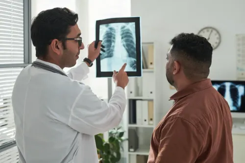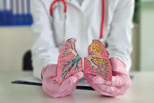
A CT scan may occasionally reveal enlarged lymph nodes or an abnormal area deep within the chest. When these structures cannot be easily sampled using conventional methods, EBUS Bronchoscopy may be considered.
EBUS stands for endobronchial ultrasound. It combines bronchoscopy with ultrasound imaging, helping doctors visualize structures adjacent to the airways and obtain samples from selected lymph nodes or lesions.
For patients in Churchgate, Mumbai, understanding how EBUS differs from conventional bronchoscopy can help when evaluating the next diagnostic step.
How Is EBUS Different From Standard Bronchoscopy?
Conventional bronchoscopy primarily allows the doctor to look inside the airways.
EBUS adds an ultrasound component. This helps identify structures beyond the visible airway wall, particularly lymph nodes located within the chest.
A needle can then be guided through the airway wall to collect samples from selected areas. This technique is commonly called EBUS-guided transbronchial needle aspiration.
Why Might EBUS Be Recommended?
The procedure can be useful when doctors need tissue or cellular samples to investigate conditions such as:
- Enlarged mediastinal lymph nodes
- Certain lung masses
- Tuberculosis
- Sarcoidosis
- Lymph node abnormalities
- Suspected lung cancer
- Other unexplained chest findings
The exact reason depends on the CT findings and clinical circumstances.
EBUS Versus More Invasive Sampling
One of the major reasons EBUS is considered is its ability to reach certain chest lymph nodes through the airways.
Traditionally, some of these areas could require more invasive surgical procedures for tissue sampling. EBUS can provide a less invasive route in appropriately selected cases.
However, it does not replace every other diagnostic procedure. The location of the abnormality, required tissue quantity, previous investigations, and suspected diagnosis all influence whether EBUS is appropriate.
What Should Patients Ask Before the Procedure?
Useful questions include:
- Why is EBUS needed in my case?
- Which lymph node or abnormality will be sampled?
- What information could the sample provide?
- Will sedation or anesthesia be used?
- Are any medicines temporarily restricted?
- What happens if the sample is inconclusive?
- When should results be expected?
Clear answers help patients understand the purpose rather than viewing EBUS as an isolated investigation.
Dr. Pujan Parikh evaluates chest imaging and respiratory findings to determine when advanced bronchoscopic assessment may contribute useful diagnostic information.
For people in Churchgate and surrounding South Mumbai locations, EBUS bronchoscopy may provide an important route to diagnosing abnormalities located near the central airways. Its value lies in obtaining targeted samples that can then be interpreted by the appropriate laboratory or pathology team.
Keywords